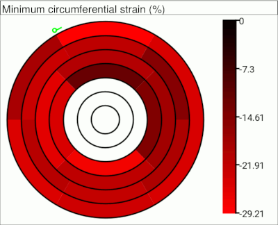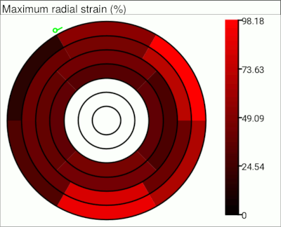
Having defined the endo- and epicardial borders at all time points, and set up the segments, you can perform cardiac strain analysis. Strain analysis uses an image registration technique to assess the movement of tissue across time-points, and is applicable to any cardiac cine sequence, whether tagged or not.
Select the "Strain" tab in the Cardiac Segment toolkit:

 button. This will start the registration process, and
after a few minutes, you will see pop-up graphs
showing the radial and circumferential strain in each segment through the
cardiac cycle, and a dialog asking
whether you want to save the results of the analysis to either a text or a PDF report:


_eRR for the radial strain
image, and _eCC for the circumferential strain. The examples
shown below were calculated from a regular (non-tagged) cine pulse sequence.
 |  |
| Radial strain image | Circumferential strain image |
|---|